Research
RESEARCH · AG CHANG
Cellular Immunology Research Group
Cytotoxic T-Cell Biology and Intercellular Communication
Molecular mechanisms in cancer, neuroinflammation and neurodegeneration

Our research group investigates the molecular mechanisms of cell–cell communication, focusing on cytotoxic T lymphocytes (CTLs), their secretory machinery, and their interactions with target cells in cancer and the nervous system.
CTLs are essential components of immune defence, eliminating infected and malignant cells through the regulated secretion of cytotoxic molecules. Our research has contributed to understanding the molecular mechanisms of cytotoxic granule trafficking, exocytosis, and endocytosis, as well as the organization and release of distinct effector cargo populations.
Beyond their classical cytotoxic functions, we investigate how cytotoxic T cells communicate with neurons and glial cells during inflammation, and how the transfer of molecular and organellar cargo influences recipient-cell survival and function. In parallel, we study Flower (CACFD1), an evolutionarily conserved protein involved in cellular fitness recognition and cell competition. Our work has uncovered a previously unrecognized mechanism by which Flower-bearing extracellular vesicles mediate fitness signalling beyond direct cell–cell contact. We investigate how Flower-dependent signalling regulates cellular survival and responses to stress in neurodegenerative and tumour-associated environments.
By integrating advanced fluorescence and electron microscopy with quantitative proteomics, transcriptomics, molecular and cellular biology, biochemistry, and functional assays, we aim to uncover how cellular secretion and intercellular cargo transfer regulate immune effector functions, determine recipient-cell fate, and influence tissue responses in cancer and neurological diseases.
1. Cytotoxic T-Cell Secretion and Effector Heterogeneity
How do cytotoxic T cells coordinate the release of their effector molecules?
We investigate the molecular organization, trafficking, and exocytosis of cytotoxic granules at the immunological synapse. Our research has contributed to understanding the origin and release of supramolecular attack particles (SMAPs), the heterogeneity of cytotoxic granules, and the coordinated secretion of granzyme B and a distinct granule-associated pool of interferon-γ (IFN-γ), termed “lytic IFN-γ.” This coordinated release enhances target-cell killing.
A major focus of our ongoing research is how CTL differentiation and physiological factors, including sex and ageing, influence secretory cargo composition and effector function.
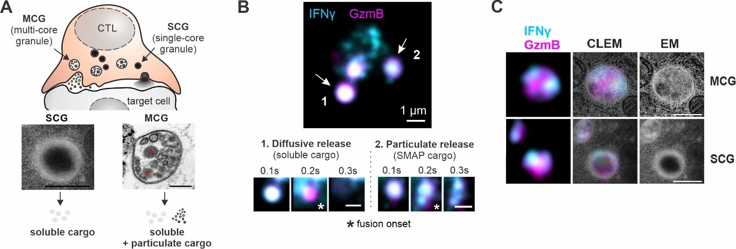
Figure 1. Cytotoxic T-cell secretion and effector heterogeneity. (A) Cytotoxic T lymphocytes (CTLs) contain morphologically distinct single-core granules (SCGs) and multi-core granules (MCGs). SCGs predominantly release soluble cargo, whereas MCGs can additionally release particulate cargo, including supramolecular attack particles (SMAPs). Red asterisks indicate SMAPs within the MCG. Scale bars: 200 nm. (B) Live-cell total internal reflection fluorescence (TIRF) microscopy reveals distinct modes of effector cargo release, including rapidly diffusing soluble cargo and particulate SMAPs that persist following granule fusion. White asterisks in (B) indicate fusion onset. Scale bars: 1 µm. (C) Correlative light and electron microscopy (CLEM) reveals heterogeneous distribution of interferon-γ (IFN-γ) and granzyme B (GzmB) among structurally distinct cytotoxic granules. Scale bars: 200 nm. MCG ultrastructure in (A) adapted from Chang et al., Nat. Commun. (2022); SCG ultrastructure in (A) and images in (B–C) adapted from Li et al., Cell. Mol. Immunol. (2026).
2. Immune–Neural Communication and Neuroinflammation
How do cytotoxic T cells interact with neural cells during inflammation?
We investigate interactions between cytotoxic T lymphocytes and cells of the central nervous system, particularly cortical neurons and glial cells, under inflammatory conditions.
Using experimental neuroinflammation models, primary neuronal cultures, brain slices, and advanced live-cell imaging, we study how cytotoxic T cells communicate with neural cells and influence their responses to inflammatory stress.
Our goal is to understand the molecular mechanisms underlying these interactions and how they contribute to neuronal injury, survival, and resilience during neuroinflammation.
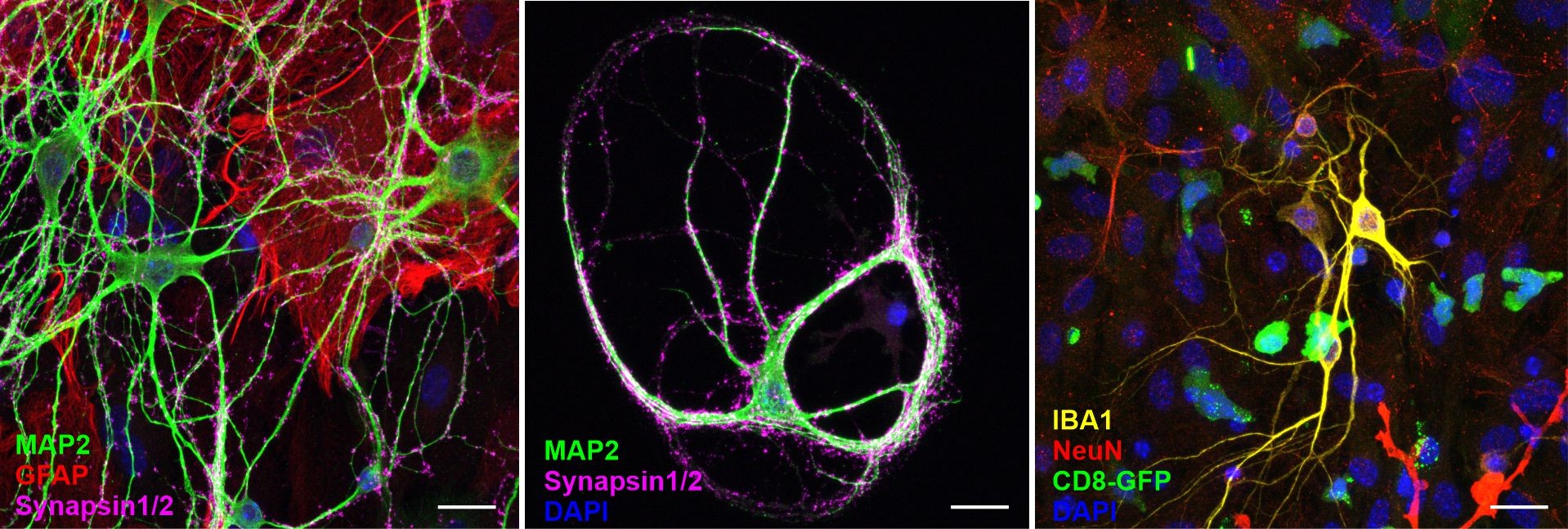
Figure 2. In vitro models for investigating immune–neural communication.
Representative fluorescence microscopy images of primary cortical neural cultures (left), autaptic neuronal cultures (middle), and T-cell–neuron co-cultures (right). Fluorescence markers are indicated in the respective panels. DAPI labels cell nuclei. Scale bars: 20 µm.
3. Cellular Fitness and Extracellular Vesicle Communication
How do cells recognize and respond to differences in cellular fitness?
Flower (CACFD1) is a protein implicated in cell competition, a biological process through which cells with different fitness states influence each other’s survival.
We investigate the molecular mechanisms underlying Flower-mediated cellular fitness selection. Our research has revealed that astrocytes release Flower-bearing extracellular vesicles that promote the elimination of less-fit recipient cells, extending fitness signalling beyond direct cell–cell contact. We also investigate how this intercellular mechanism operates alongside Flower-dependent, cell-intrinsic protection to regulate astrocyte survival.
We study how Flower-mediated fitness signalling influences astrocyte responses to amyloid-β stress and neuronal protection, and explore its role in cellular interactions within tumour-associated environments.
By integrating molecular mechanisms with disease-relevant models, we aim to understand how cellular fitness signalling shapes tissue responses to inflammation and stress.
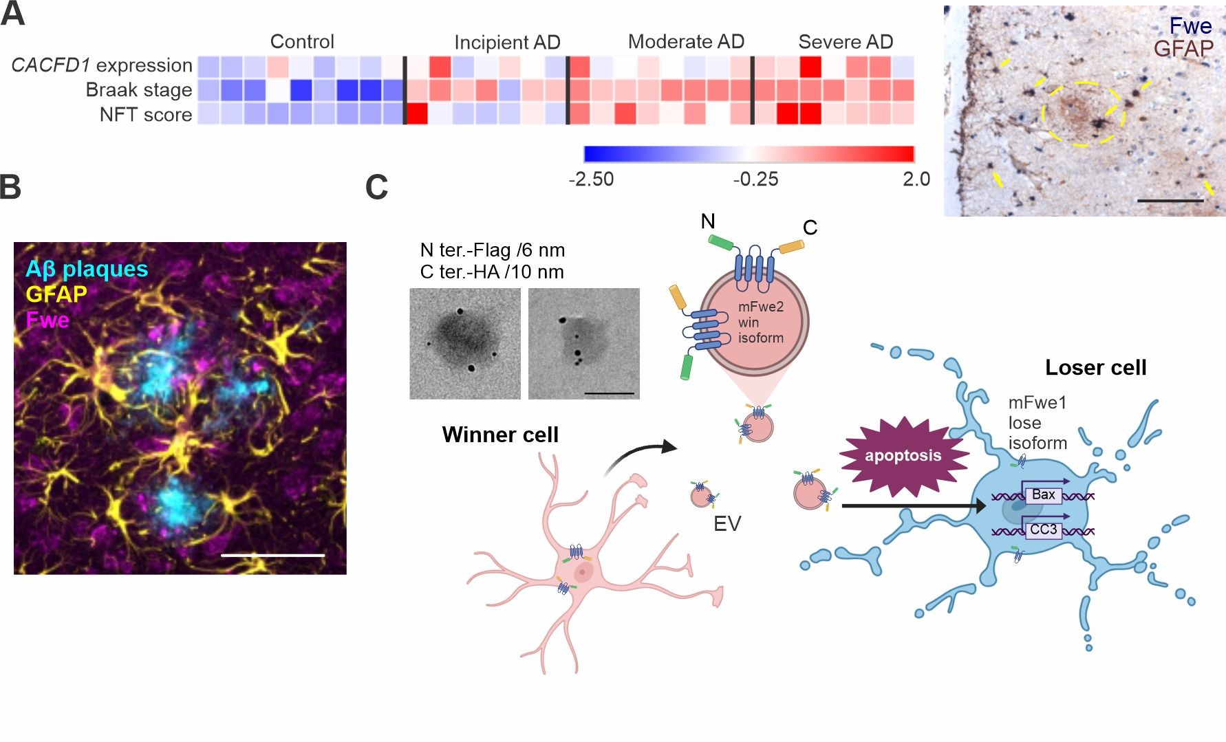
Figure 3. Flower-dependent cellular fitness and extracellular vesicle communication in neurodegeneration. (A) Heatmap showing CACFD1 (Flower) expression, Braak stage and neurofibrillary tangle (NFT) score in human Alzheimer’s disease (AD) brain samples and controls from dataset GSE1297. CACFD1 expression is shown as normalized relative expression levels, with blue indicating lower expression and red indicating higher expression. Representative immunohistochemical staining shows Flower (Fwe) and the astrocyte marker GFAP in post-mortem human AD brain tissue. Arrows indicate Flower-positive astrocytes; the dashed circle marks a plaque. Scale bar: 50 µm. (B) Confocal imaging showing enrichment of Flower-positive astrocytes around amyloid-β plaques in APP-transgenic mouse cortex. Scale bar: 50 µm. (C) Proposed model of Flower-mediated cellular fitness signalling, in which winner astrocytes release Flower-bearing extracellular vesicles (EVs) that induce apoptosis in less-fit recipient cells. Immunogold electron microscopy demonstrates surface accessibility of both the N- and C-terminal epitopes of tagged Flower on EVs. Scale bar: 100 nm. Images and model adapted from Tu et al., EMBO Reports (2026).
Research Networks
Our group contributes to the ERC Synergy ATTACK consortium (https://supramolecular-attack-particles.eu/), the Center for Gender-specific Biology and Medicine (CGBM), and PharmaScienceHub (PSH) (https://pharmasciencehub.com/).